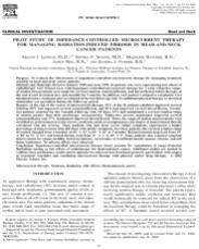
To evaluate the effectiveness of impedance-controlled microcurrent therapy for managing treatment sequelae in head-and-neck cancer patients. Methods and Materials: Between January 1998 and June 1999, 26 patients who were experiencing late effects of radiotherapy were treated b.i.d. with impedance-controlled microcurrent therapy for 1 week. Objective range of-motion measurements were made for cervical rotation, extension/flexion, and lateral flexion before therapy, at the end of each treatment day, and monthly for 3 months. In addition, each patient’s subjective complaints were tabulated before treatment and reevaluated at the last follow-up visit. No additional physical therapy or electrical stimulation was permitted during the follow-up period. Results: At the end of the course of microcurrent therapy, 92% of the 26 patients exhibited improved cervical rotation, 85% had improved cervical extension/flexion, and 81% had improved cervical lateral flexion. Twenty two patients returned for the 3-month follow-up visit. Of these, 91% had maintained a cervical rotation range of motion greater than their pretherapy measurements. Eighty-two percent maintained improved cervical extension/flexion and 77% maintained improved lateral flexion. When the range-of-motion measurements were stratified by pretreatment severity (severe, moderate, mild, or asymptomatic), the degree of improvement directly correlated with the severity. Thus, patients who had more severe initial symptoms experienced a higher percentage of improvement than did those with milder symptoms. For these patients, the cervical rotation range of motion changed from a baseline of 59° 12° to 83° 14° at 3 months; flexion/extension improved from 47° 10° to 73° 13°; and lateral flexion went from 31° 7° to 48° 9°. Some patients also reported symptom improvement for tongue mobility, facial asymmetry, xerostomia, cervical/facial muscle spasms, trismus, and soft tissue tenderness. No adverse effects were observed. Conclusion: Impedance-controlled microcurrent therapy shows promise for remediation of range-of-motion limitations arising as late effects of radiotherapy for head-and-neck cancer. Additional studies are needed to validate these preliminary results and to optimize the microcurrent treatment protocol, particularly with respect to treatment schedules and combining microcurrent therapy with physical and/or drug therapy. © 2002 Elsevier Science Inc. Microcurrent therapy, Neutrons, Radiation, Side effects, Head-and-neck cancer. INTRODUCTION As aggressive therapy with combination surgery, chem studies (2–4) from the 1980s suggesting that microcurrent therapy was effective for treating RT sequelae, but these studies lacked adequate statistics and did not include follow-up information on the long-term effectiveness. The reports also lacked information on the specific treatment instruments and precise treatment protocols used. This pilot study was designed to determine whether the suggested efficacy would be observed in a series of patients treated using a well-specified protocol. METHODS AND MATERIALS Twenty-six head-and-neck cancer patients who had completed RT and were experiencing tissue discomfort or limitations caused by fibrosis participated in the study. Because this was a pilot study to determine the efficacy of a new use of a standard therapeutic technique, it was important that all participants have quantifiable symptoms with no expectation of resolution without intervention. Hence, patients experiencing documented progressive fibrosis were targeted. The staff made objective range-of-motion measurements, and subjective complaints were solicited from the patients. The procedure and its possible lack of benefit were explained to the patients before they signed a document indicating informed consent. The Provena Saint Joseph Hospital Institutional Review Board approved the protocol. Selection of study subjects Eligible patients had finished either photon or neutron therapy at least 6 months before entering the study and had no evidence of disease. They had mental alertness sufficient to understand, evaluate, and consent to the protocol, which included the availability for b.i.d. treatments daily for 1 week and the ability to return for scheduled follow-up visits. Exclusion criteria included the use of a pacemaker, use of calcium-channel blocker drugs, pregnancy, and a life expectancy of 6 months. Individuals who were unable to abstain from physical therapy to the affected area, routine use of antiinflammatory steroids, or nonsteroidal antiinflammatory drugs during the treatment and follow-up period were also excluded. Table 1 summarizes the baseline characteristics of the participants. Choice of microcurrent technique and schedule The use of electrical stimulation for pain relief is well established in physical therapy centers. Many commercial electrical stimulation devices are available, most of which are commonly referred to as transcutaneous electrical nerve stimulation units. Typical units emit electrical pulses with alternating positive and negative polarities in the 10–500- kHz range and currents in the milliampere range. Microcurrent units are often incorrectly referred to as transcutaneous electrical nerve stimulation units, but microcurrent units deliver lower currents (microampere range) and lower frequencies (0.5 to several hundred hertz). In general, units using higher current and frequencies are more effective at blocking acute pain, but the pain relief is not lasting. Microcurrent therapy using lower frequencies requires longer treatment times to achieve pain relief, but the relief can endure for many hours after the treatment has terminated (5). Because the patients targeted for this study were experiencing chronic rather than acute symptoms, a microcurrent device was selected. The costs of microcurrent devices range from several hundred to thousands of dollars. Some fraction of the cost is related to packaging, but most of it is associated with the degree of sophistication of the electronic circuits. It is well known that the body’s impedance changes when electrical current passes through it. The more sophisticated devices contain circuitry that monitors impedance and adjusts the output current to compensate for changes. These devices also deliver fast rise time pulses that can affect voltagesensitive sodium and calcium ion channels (6). The ElectroMyopulse and Electro-Acuscope instruments (Biomedical Design Instruments, Burbank, CA) chosen for this study deliver impedance-controlled, fast rise time pulses. Their retail price is about $8500 each. Electrotherapy treatments are reimbursable under established billing codes. Typical charges to a patient are $40–50 per 15-min treatment. However, patients in this study were not charged for the therapy. Physical therapists use microcurrent therapy in a variety of ways, often in combination with massage, heat, and physical manipulation. Treatment schedules are not standardized, but are driven by insurance payment schedules and the patients’ personal schedules. The treatment schedule for this study was established after informal discussions with a few physical therapists who had extensive experience using the Electro-Myopulse and Electro-Acuscope instruments for treating a variety of physical complaints. All agreed that noticeable improvement could be obtained most quickly if the patient were treated b.i.d. for 3 days. All agreed that lasting improvement tended to require several treatments per month for about 6 months and that some conditions could resolve completely if this long-term treatment schedule were followed, particularly if therapy started soon after the injury or symptom occurred. Given the adTable 1. Baseline characteristics of 26 patients in the pilot study Fast neutrons Photons Neutrons and photons Gender (n) Male 3 9 2 Female 5 4 3 Race (n) White 8 13 3 Black 0 0 2 Age (y) 52 15 56 9.3 63 15 Radiation dose (Gy) 20.8 0.8 64 8.3 20.3 0.1 (n) 36 25 ( ) Time from RT to start of therapy (mo) 67 61 30 27 42 38 Data presented as the average standard deviation, unless otherwise noted. Abbreviations: RT radiotherapy; n neutrons; photons. 24 I. J. Radiation Oncology ● Biology ● Physics Volume 54, Number 1, 2002 vanced fibrosis of many of the study patients, it was decided to administer microcurrent treatments b.i.d. for 5 days and simply observe whether this therapy had any effect on severely fibrotic tissue. Any observed improvements were not expected to be lasting, because no follow-up treatments at more spread-out intervals were scheduled. Until measurable evidence of the treatment’s effectiveness was observed, it did not seem reasonable to commit resources to a longterm treatment schedule. Objective measurement techniques As shown in Fig. 1, cervical rotation, extension/flexion, and lateral flexion were measured using two large protractors mounted in perpendicular planes. An elastic band with Velcro attachments was secured to the patient’s head to permit the placement of a small laser that pointed to degree markings on circular scales used to measure range of motion in degrees. This laser was positioned relative to the points about which the patient’s head pivots during rotation, exFig. 1. Patient positioned at vertex of two mutually perpendicular protractors used to measure cervical range of motion. Fig. 2. Laser affixed to the patient’s head measures left–right cervical rotation. Impedance-controlled microcurrent therapy for RT-induced fibrosis in head-and-neck cancer ● A. J. LENNOX et al. 25 tension/flexion, and lateral flexion. Stationary lasers were used to position the patient so that the movable laser was on a line that intersected the vertex of the large protractors. Figures 2 through 4 illustrate the setup for each angular measurement. Day-to-day patient positioning accuracy was 0.25 cm, which is small compared with the protractors’ 112-cm radius. This choice of scale minimized the effect of day-to-day errors in positioning the patient’s center of rotation at the vertex of the scale. For each patient, the pretreatment data were used to classify each range of motion as asymptomatic or mildly, moderately, or severely limiting. If a patient’s range was within 90% of the optimal range for a healthy young person, that patient was classified as asymptomatic for that measurement. Ranges between 70% and 90% of optimum were designated mildly limiting, and those of 50–70% were moderately limiting. Ranges 50% of optimum were considered severely limiting. By assigning a value of 0 to Fig. 3. Cervical extension/flexion measured using a laser affixed to the side of the head. Fig. 4. Cervical lateral flexion measured using a laser affixed to the forehead. 26 I. J. Radiation Oncology ● Biology ● Physics Volume 54, Number 1, 2002 Table 2. Patient characteristics listed in order of greatest to least severe radiation-induced range-of-motion limitations before impedance-controlled microcurrent therapy Severity RT site Dose (Gy) Radiation Pathologic features Stage Other therapy 9 Left thyroid Bilateral neck Supraclavicular nodes 66 66 e e Medullary carcinoma T4N1bM0/Stage 3 Surgery 9 Oropharynx 63 e Squamous cell T1N2bM0 Surgery Bilateral neck Supraclavicular nodes 50.4 e 9 Left tonsil Bilateral neck Supraclavicular nodes 74.4\